Imagej Quantify Fluorescence Intensity
Make sure you have area integrated intensity and mean grey value selected the rest can be ignored. It actually subtracts intensity from each pixel and results in an image that is less intense visibly than the max projection.

Fluorescence Intensity Measurement Of The Roi With Imagej A

Imagej Create A Color Intensity Map

Quantitative Analysis Of Histological Staining And
Measuring cell fluorescence using imagej image j can be downloaded for free from here here is a very simple guide for determining the level of fluorescence in a given region eg nucleus 1.
Imagej quantify fluorescence intensity. Basic intensity quantification with imagej pretty pictures are nice but many times we need to turn our images into quantifiable data. After that it will find the minimum intensity in the bleached roi and fit the recovery with this point in mind. Now select measure from the analyze menu.
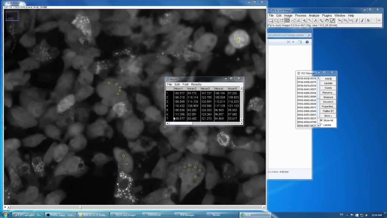
There are a number of different ways to get intensity information from images using the base package of imagej no plugins required. How can i measure fluorescence intensity and distribution in imagej. Open the roi manager.
Hi all i am new to imagej and i need to quantify immunofluorescence intensity and distribution of protein in the image. For calcium imaging you want average intensity within the cell so you should check mean gray value for your graph you probably want to calculate ff0 mean fluorescence at time t divided by the mean at time 0 for each cell. You should now see a popup box with a stack of values for that first cell.
Imagej is useful for getting information from images including pixel intensity. Frap fluorescence recovery after photobleaching analysis. The frap profiler plugin will analyze the intensity of a bleached roi over time and normalize it against the intensity of the whole cell.
Notice that your rectangular rois include area both inside and outside the cell. Select the cell of interest using any of the drawingselection tools ie. Measuring cell fluorescence using imagej.
Using imagej to measure cell number and cross sectional area of confocal images. Imagej how to measure mean fluorescence intensity over timelapse image stack condensed duration. You will then need to select an area corresponding to a single celegans within the image and that you can easily do with imagejfiji and then measure the fluorescence intensity.
I am also trying to quantify fluorescence intensity in a z stack but i noticed that the sum projection method in imagej is not a true sum of pixel intensity in the z direction for any pixel xy. Rectangle circle polygon or freeform 2.
Imagej How To Measure Mean Fluorescence Intensity Over Timelapse Image Stack Condensed
Analyze Menu
Plos One Attenuation Of Inflammatory Mediators Tnf α And

Publicquantifyingcolorintensity Wiki It Support
Stowers Imagej Plugins

Spot Intensity Analysis Imagej
Analyzing Gels And Western Blots With Imagej Lukemillerorg

Measuring Intensity Using Imagej Stack Overflow
Plos One Regulation Of Fatty Acid Oxidation In Mouse

Quantifying Microglia Morphology From Photomicrographs Of
Plos One Loss Of Epithelial Markers Is An Early Event In

About The Quantification Of Puncta Like Staining

How To Take Area Measurements Of Cells Using Imagej

Count Nuclear Foci Imagej Duke Light Microscopy Core

Selective And Uncoupled Role Of Substrate Elasticity In The
Quantitative In Situ Hybridization With Enhanced Sensitivity
Plos One Sumoylation Is Required For Glycine Induced
Analyzing Fluorescence Microscopy Images With Imagej

Publicquantifyingcolorintensity Wiki It Support